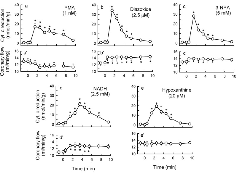
Figure 11.
Outflow of reduced cytochrome c (a–e) and coronary flow (a'–e') in guinea pig hearts stimulated for 10 min with 1 nM PMA (a,a'), 2.5 μM diazoxide (b,b'), 5 mM 3-nitr-propionic acid (3-NPA, c,c'), 2.5 mM NADH, d,d') and 20 μM hypoxanthine (e,e'). The agents were introduced into the perfusate at time 0. Values are means ± SEM of 5–6 experiments. * p < 0.05 vs. basal measurements.

