Figure 1.
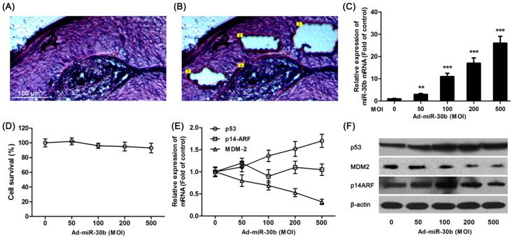
Overexpression of miR-30b regulated p53 expression. (A) Hematoxylin and eosin (H&E) staining of laryngeal carcinoma sections; (B) Using LCM (laser capture microdissection), sections were divided into center area (Area 4) paracancerous (paraneoplastic distance <5 mm, Area 1–3) and surgical margins (paraneoplastic distance >10 mm, not shown) by LCM. Scale bar = 100 μm and referred to (A) and (B) panels; (C) After HEp-2 cells were incubated with miR-30-lentivirus vector for 16 h in different multiplicity of infection (MOI), the expression of miR-30b was determined using qRT-PCR. ** p < 0.01, *** p < 0.001 compared to control group, n = 6; (D) After infected by miR-30-lentivirus vector for 16 h, HEp-2 cell viability was detected by 3-(4,5-dimethyl-2-thiazolyl)-2,5-diphenyl-2-H-tetrazolium bromide (MTT) assay; (E) After infected, the mRNA expression level of p53, MDM-2 and p14-ARF were investigated using qRT-PCR; and (F) The expression of p53, MDM-2 and p14-ARF in protein level were detected by western blot after infected. All experiments were repeated at least three times.

