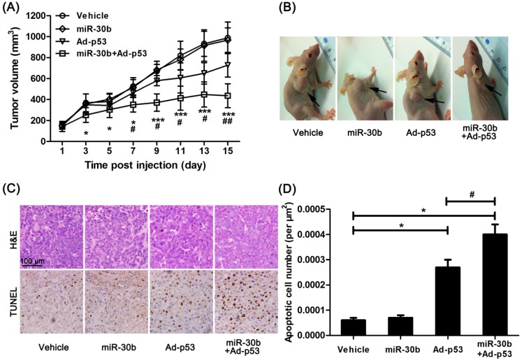
Figure 3.
Overexpression of miR-30b improved the anti-tumor effect of Ad-p53 in vivo. (A) After Ad-p53 injection, tumor volume was calculated every two days, * p < 0.05, *** p < 0.005 compared to normal HEp-2 cell-implanted nude mice, # p < 0.05, ## p < 0.01 compared to Ad-p53-treated implanted mice, n = 8; (B) After 15 days injection, the tumor in nude mice was pictured, the black arrow in the representative figure indicating tumor position; (C) After the experiment, the tumor tissue was removed and made into sections. H&E and TdT-mediated dUTP nick end labeling (TUNEL) staining were performed. Brown showed TUNEL-positive nuclei, and all nuclei were stained with hematoxylin. Scale bar = 100 μm and referred to all panels; and (D) Apoptotic cell number was calculated as TUNEL-positive number divided by total cells. * p < 0.05 compared to normal HEp-2 cell-implanted nude mice, # p < 0.05 compared to Ad-p53-treated implanted mice, n = 8.

