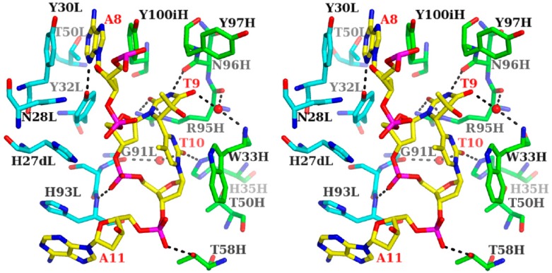
Figure 5.
Stereo drawing of the antigen-binding site of the 64M-5 Fab—ds-(6-4) DNA complex (PDB ID: 3VW3). The AT(6-4)TA nucleotide of ds-(6-4) DNA is shown in yellow and labeled in red. The heavy chain of Fab is drawn in green, and the light chain in cyan. Hydrogen bonds are shown in broken lines. T9 and T10 correspond to the T(6-4)T segment. Water molecules are drawn with spheres.

