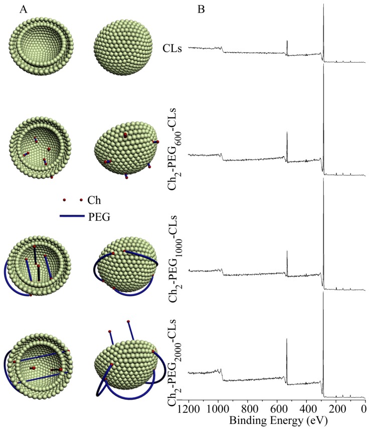
Figure 4.
Schematic diagram and XPS analysis of Ch2-PEGn-CLs. (A) Ch segments of Ch2-PEGn anchored into the lipid bilayer of CLs. Due to the short PEG chain, Ch2-PEG600 did not effectively shield the charge of CLs. Ch2-PEG1000 and Ch2-PEG2000 decreased the positive charge by covering the surface of CLs. With longer PEG chains, Ch2-PEG2000 compressed the liposomal particle and therefore Ch2-PEG2000-CLs showed a smaller particle size; (B) XPS analysis demonstrated that more PEG segments were located on the surface of liposomes when PEG molecular weight increased.

