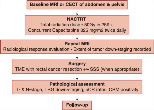
Figure 1.

Flow chart depicting the conduct of the study. A baseline computed tomography was used in some patients who did not have baseline magnetic resonance imaging (MRI – Magnetic resonance imaging; CECT – Contrast-enhanced computed tomography; pCR – Pathological complete response; TME – Total mesorectal excision; SSS – Sphincter saving surgery; CRM – Circumferential resection margin; T – Tumor; N – Node)
