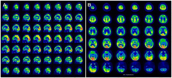
Figure 4.

PET imaging results: brain. Remarkable abnormalities are seen in the basal brain metabolic rate in a patient with CTX. PET reveals hypometabolism in cerebral lobes (especially in the frontal and temporal lobes) in sagittal section (A), and in axial section (B).
