Figure 7.

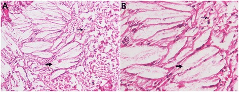
Histology: tendon. HE staining of the tendon masses reveals accumulation of xanthoma cells (fine arrows) and dispersed lipid crystal clefts (coarse arrows). A, 100×; B, 200 × .

Histology: tendon. HE staining of the tendon masses reveals accumulation of xanthoma cells (fine arrows) and dispersed lipid crystal clefts (coarse arrows). A, 100×; B, 200 × .