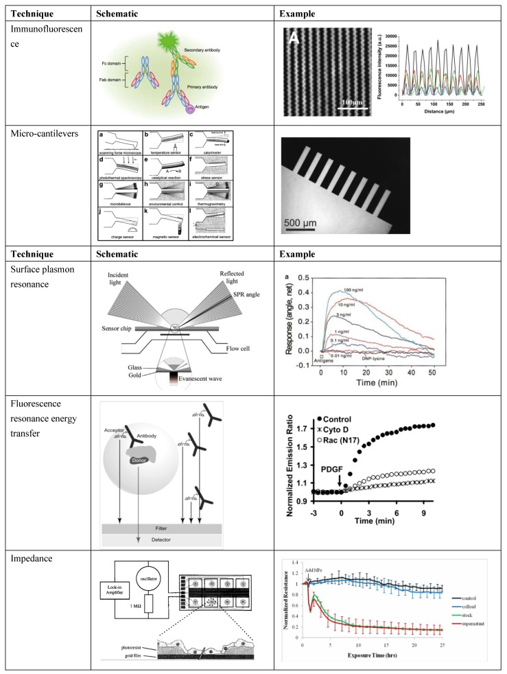
Figure 1.
Select biosensor transducer schematics and examples of use. Immunofluorescence schematic [30] and example involving sensing in microgrooves [24]; microcantilevers schematic [6] and example of a fabricated microcantilever array [6]; SPR schematic [22] and example involving detection of concentration of a molecule over time [31]; FRET schematic [32] and example [33] of signal in perturbed and normal cells; impedance schematic [34] and example of signal in control and cells with a toxin [35] (Adapted by permission from Macmillan Publishers Ltd.: J. Invest. Dermatol. [30], ©2013; reused with permission from Elsevier [6,22,24,31,32,34]; with permission ©2008 by the National Academy of Sciences [33]; with permission from Inderscience Ltd. [35], ©Inderscience 2011, respectively).

