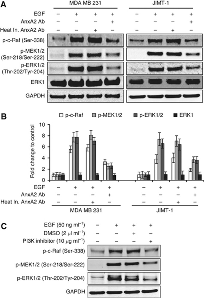
Figure 7.
AnxA2 antibody inhibits the EGF-induced ERK pathway in MDA-MB-231 and JIMT-1 cells. (A) After 12 h serum starvation, cells were treated with or without EGF (50 ng ml−1) for 20 min after 2 h of heat inactivated AnxA2 (D1/274.5) antibody (2 μg ml−1) AnxA2 (D1/274.5) antibody (2 μg ml−1) pretreatment. Western blot analyses were carried out with indicated antibodies. (B) A bar graph showing densitometric analysis of bands of protein used in (A). Each bar represents the mean±s.e. of three independent experiments. (C) After 12 h of serum starvation, JIMT-1 cells were treated with or without EGF (50 ng ml−1) for 20 min after 2 h of PI3K inhibitor (10 μg ml−1) pretreatment. The cell lysate was analysed for p-c-Raf (Ser-338), p-MEK (Ser-218/Ser-222), p-ERK (Thr-202/Tyr-204) by western blotting.

