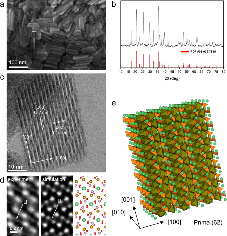
Figure 1.
Structural characterization of the LiFePO4 NCs. (a) SEM image of several nanoplatelets. (b) XRD pattern consistent with olivine-type LiFePO4 (triphylite, PDF #01-072-7845). (c) HRTEM image of a single platelet close to [010] zone axis projection. (d) Enlarged view from a [010] orientation (left) after averaging several regions of the platelet, along with simulated image (center) and sketch of atomic columns in projection (right); (e) Atomistic sketch of a LiFePO4 platelet. FeO6 octahedra are shown in brown, PO4 tetrahedra in orange, and Li ions in green.

