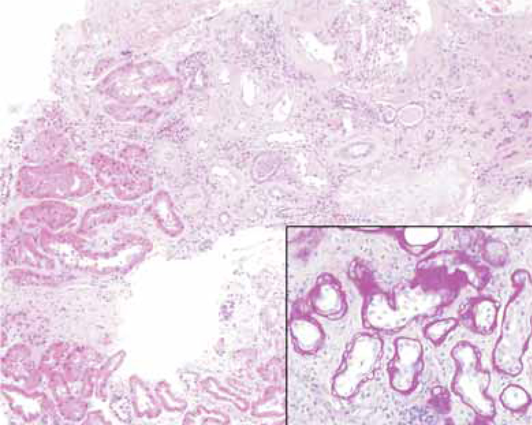
Figure 3.
Renal biopsy (Hematoxylin and eosin stain). Kidney biopsy of patient AII6 reveals patchy interstitial fibrosis accompanied with focal chronic inflammatory infiltrate, and focal tubular atrophy with markedly thickened and duplicated basement membranes (insert, periodic acid-Schiff). Focal proximal tubules showed hypertrophy. Only one glomerulus was present and showed periglomerular fibrosis but no other alterations by light microscopy.

