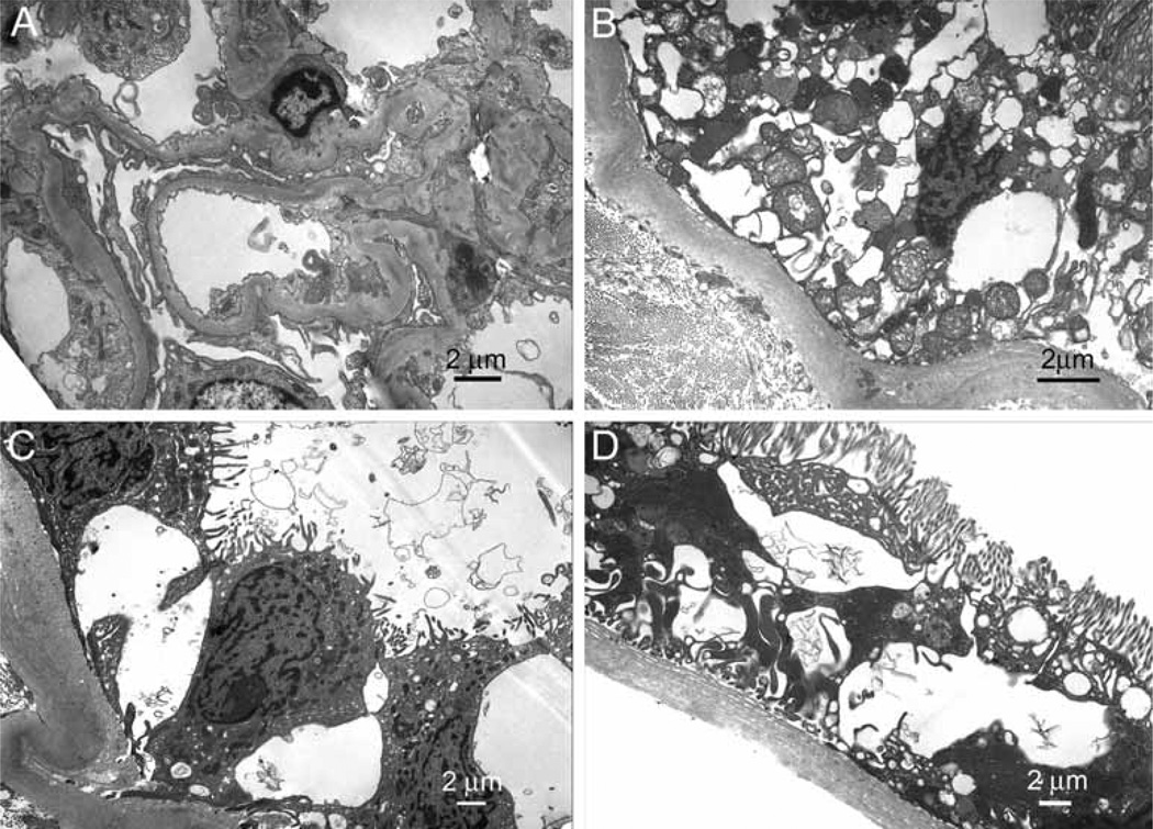
Figure 4.
Ultrastructural examination of kidney biopsy specimen from patient AII6. A: Glomerulus with segmentally thickened and homogenous basement membranes. Segmental podocyte foot process effacement was present. B: Epithelial cells of proximal tubule demonstrating distention of basal membrane invaginations and thickening of tubular basement membrane. C: Distal tubule with marked distention of the intercellular spaces between epithelial cells and thickening of tubular basement membrane. D: Proximal tubule showing epithelial cell atrophy without any significant reduction of the brush border. Thickening of tubular basement membrane and distention of basal membrane invaginations are also present.

