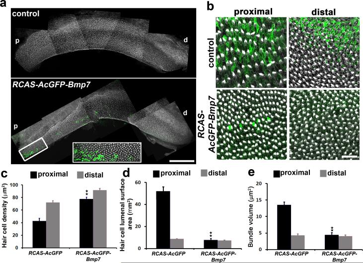
Figure 6. Over-expression of Bmp7 in ovo alters positional identity along the tonotopic axis.
(a) Low magnification montage images of E14 BPs electroporated with either RCAS-AcGFP(control) orRCAS-AcGFPBmp7at E2.5. Images show BPs labeled with phalloidin (white) to visualize stereociliary bundles and anti-GFP (green) to visualize infected cells. Electroporation does not alter the normal morphology of the developing BP. (b) High magnification images of the indicated regions of the BP from control (top) and RCAS-AcGFP-Bmp7(bottom) samples labeled as in a. Expression of Bmp7 in the proximal region of the BP induces a marked increase in hair cell density and decrease in lumenal surface area. (c)Quantification of changes in mean hair cell density in proximal (black bars) and distal (grey bars) regions of the BP following electroporation with RCAS-ACFGP(control) or RCAS-AcGFP-Bmp7. Proximal Hair cell density is significantly increased in the presence of Bmp7. (d)Quantification of mean hair cell lumenal surface area following electroporation. Expression of Bmp7 induces a significant decrease in proximal hair cell surface areas.(e) Mean stereociliary bundle area for hair cells located in proximal (black bars) or distal (grey bars) regions of the BP following electroporation with either RCAS-AcGFP or RCAS-AcGFP-Bmp7.Bmp7 induces significantly decreased bundle area in proximal hair cells. Data are mean ±sem. **: p< 0.05 based on Student's t-test. Scale bars (a = 200 μm, b = 50 μm). For controls n = 4 and for Bmp7 n = 7.

