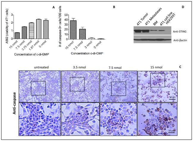
Figure 2. High doses of c-di-GMP killed 4T1 tumor cells directly and activated caspase-3.
4T1 tumor cells were incubated with various doses of c-di-GMP for 24hours. The viability of the 4T1 tumor cells was assayed by MTT assay (A), or the number of 4T1 tumor cells with caspase-3 activation was quantified by immunohistochemistry (IHC) (B). The results shown here is the average of three independent experiments. The number of caspase-3-positive cells was counted per 100 cells. The error bars represent the SEM. An example of caspase-3 activation is shown by IHC (C). Also the expression of STING was analyzed in the primary tumor, metastases and the 4T1 cell line by western blotting (D). Bone marrow (BM) cells and HEK293 were used as a positive and negative control, respectively.

