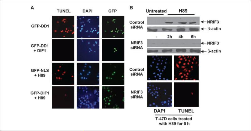
Figure 5.
DIF-1 rescues breast cancer cells from DD1-mediated and H89-mediated apoptosis. A, T-47D cells were transfected with 100 ng pLPC-DIF-1. Other cells were transfected with control pLPC vector. Twenty-four hours later, the cells were transfected with 50 ng of GFP-DD1 vector. Five hours later, cells were analyzed for apoptosis by TUNEL assay. Shown are represented fields. Expression of DIF-1 decreased DD1-mediated apoptosis by over 90%. For the H89 study, T-47D cells were transfected with GFP-DIF-1 vector (100 ng) or with a vector expressing GFP containing a nuclear localization signal (GFP-NLS; 75 ng). Twenty-four hours later, the cells received 200 nmol/L of H89, a PKA inhibitor. Five hours later, the cells were examined for apoptosis by TUNEL assay. Shown are representative fields. Expression of DIF-1 resulted in a >90% reduction in apoptosis mediated by H89. B, H89 mediates apoptosis in breast cancer cells through NRIF3. T-47D cells were treated with a NRIF3 siRNA (25 nmol/L) or with a control siRNA (25 nmol/L). Thirty hours later, the cells received 200 nmol/L H89. Cells were harvested for NRIF3 expression by Western blotting 2, 4, and 6 h after addition of H89. Cells were also examined for apoptosis by TUNEL assay 5 h after H89 incubation. H89 incubation results in a rapid increase in NRIF3. NRIF3 siRNA blocked the rapid H89-mediated increase in NRIF3 expression, as well as apoptosis. Supplementary Fig. S5 indicates that this rapid increase in NRIF3 occurs at the mRNA level.

