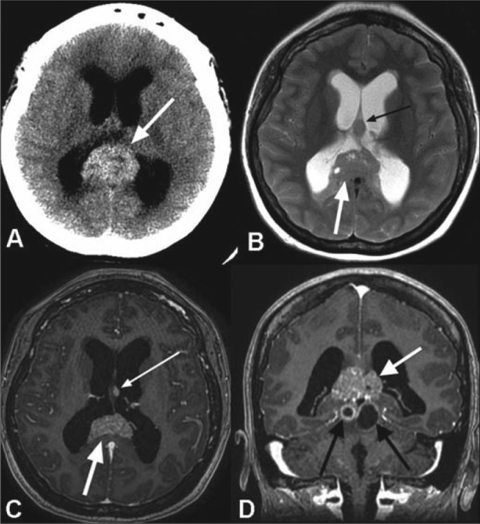
Figure 1.

(a) Axial noncontrast CT image demonstrates a pineal region mass (white arrow) with intrinsic hyperdensity and associated obstructive hydrocephalus. (b) Axial T2-weighted, (c) axial, and (d) coronal postcontrast T1-weighted MR images reveal a T2 signal isointense to gray matter with intermixed cystic foci (white arrow in b) and avid enhancement following contrast administration (large white arrows in c and d). A separate T2 hypointense (small black arrow in b) and enhancing (small white arrow in c) nodule is seen at the interface of the septum pellucidum and left forniceal body, which suggests local cerebrospinal fluid dissemination of tumor. Additional, peripherally enhancing cystic foci are present within the midbrain tectum (black arrows in d).
