
Figure 3.
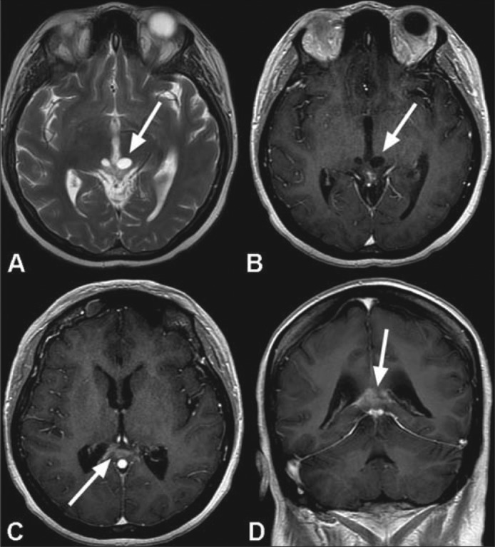
MR images following radiotherapy show a marked improvement in the appearance of the tumor bed. Near complete resolution of the large mass in the pineal fossa is seen with minimal residual enhancement along the margin of the splenium of the corpus callosum (arrows in c and d). There is considerable reduction in the extent of the previously seen peripheral enhancement involving the cystic components within the tectum of the midbrain (arrows in a and b).