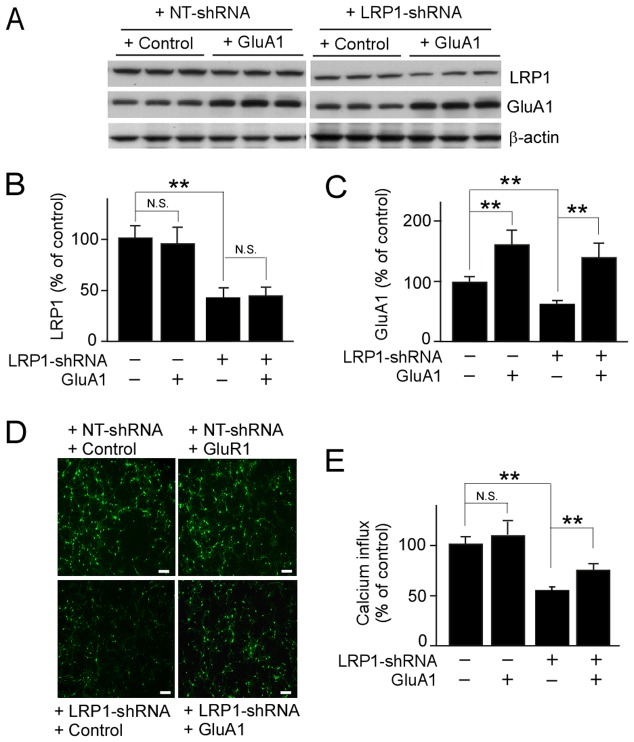
Figure 4. LRP1-knockdown suppresses GluA1-mediated calcium influx in neurons.
Primary mouse neurons were first infected with lentivirus carrying control vector or GluA1 plasmid, and then with lentivirus carrying NT-shRNA or LRP1-shRNA (A). Expression levels of LRP1 (B) and GluA1 (C) were detected by Western blot. (D) Calcium influx detected with the fluorescence microplate reader using Fluo-4 AM as a fluorescent indicator of intracellular calcium concentration in neurons after stimulation of AMPA in the presence of NMDAR antagonist. The scale bar represents 200 µm. (E) Calcium fluorescence intensities were measured with the excitation and emission wavelengths set at 494 and 535 nm, respectively. The data are plotted as mean ± SD (n = 3). N.S., Not significant; **, p<0.01.

