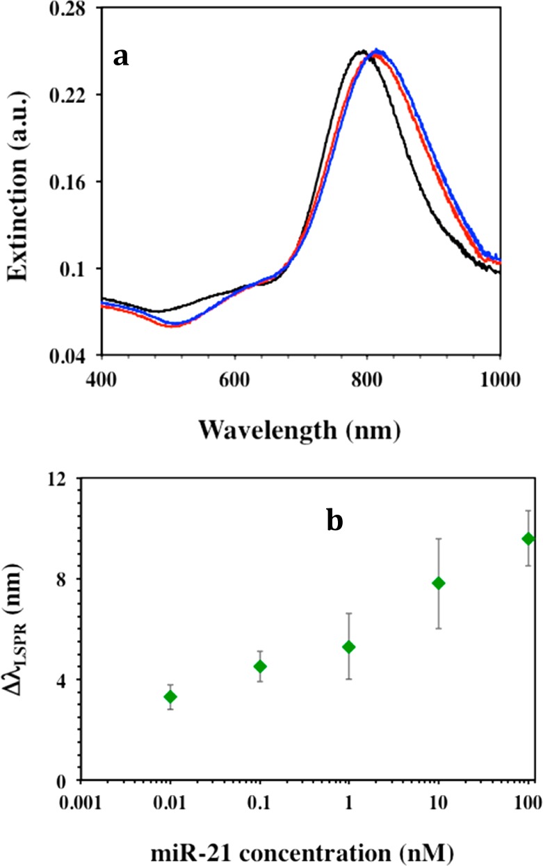
Figure 3.

Determining the optimum detection condition. (a) UV–visible extinction spectra monitoring the LSPR dipole peak (λLSPR) of gold nanoprisms attached to silanized glass substrate before (black, λLSPR = 796 nm) and after (red, λLSPR = 811 nm) functionalization with 1 μM of HS-C6-ssDNA-21 without PEG6-SH spacers and after incubation in 100 nM miR-21 solution in 40% human plasma (blue, λLSPR = 822 nm). (b) Average ΔλLSPR of these HS-C6-ssDNA-21 functionalized gold nanoprisms upon hybridization with different miR-21 concentrations in 40% human plasma. The ΔλLSPR were calculated by taking the difference between the λLSPR peak position of the nanoprisms after and before the incubation with miR-21 in PBS buffer.
