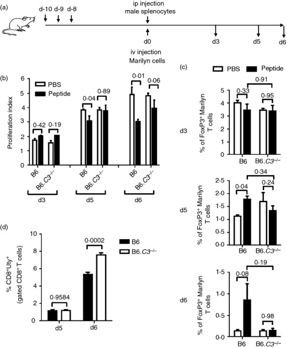
Figure 4.

Induction of antigen-specific regulatory T (Treg) cells in peptide-treated mice after immunization with male cells. Ten days after intranasal (i.n.) treatment with three doses of peptide (100 μg), female B6 wild-type (WT) or B6.C3–/– mice (three or four animals per group) were challenged intraperitoneally with 5 × 10⁶ male splenocytes. The antigen-specific CD4 T-cell response was monitored by infusing 2 × 10⁶ CFSE-labelled Marilyn CD90.1 CD4+ CD25− T cells intravenously. Three, 5 and 6 days after the challenge, splenocytes were recovered and the proliferation of Marilyn T cells (gated as CD4+ CD90.1+) was analysed by flow cytometry (a) Experimental schedule. (b) Proliferation indices of Marilyn T cells. (c) Percentage of Foxp3+ Marilyn T cells. Results from mice treated with PBS (open column) and peptide (filled column) are shown. Bars indicate SEM; P-values of unpaired t-test are indicated. (d) Five and 6 days after the challenge, splenocytes were recovered and analysed for HY-specific CD8 T-cell responses (HY-DbUty dextramer+ CD8 T cells) by flow cytometry. Bars indicate SEM. P-values of unpaired t-test are indicated.
