Figure 3.

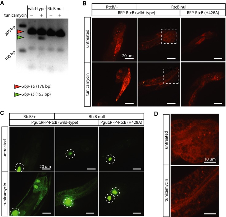
RtcB ligates the xbp-1 mRNA
A RT–PCR using primers that detect both xbp-1U and xbp-1S. In wild-type, xbp-1S is observed after 5 h of tunicamycin treatment. In RtcB nulls, xbp-1S is not detected.
B Expression of RFP-tagged wild-type RtcB and RFP-tagged inactive RtcB (H428A) in the intestine of RtcB/+ controls or RtcB mutant animals. Both wild-type and inactive RtcB are robustly expressed. Dashed boxes are detailed in (D).
C Same animals as (B), visualizing GFP expression from the Phsp-4::GFP reporter under normal conditions (top panels) and after induction of ER stress (bottom panels). Expression of inactive RtcB (H428A) fails to rescue GFP induction in RtcB null mutants. Dotted circles indicate GFP expression in coelomocytes from the injection marker. Scale bars, 20 μm.
D RFP-tagged RtcB is diffusely localized in the cytoplasm and nucleus. Images correspond to dotted squares in panel (B). Scale bars, 10 μm.
