
Figure 1.
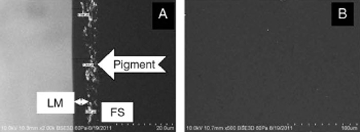
Scanning electron microscopic (SEM) images of 1DAD. Cross-sectional view at 2,000× magnification showing pigment enclosed within the lens matrix (LM) below the front lens surface (A) and an image of the lens front surface at 500× magnification showing no pigment particles (B). FS: front surface. 1DAD: 1-DAY ACUVUE DEFINE.