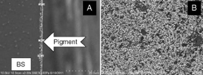
Figure 3.

Scanning electron microscopic (SEM) images of DM2. Cross-sectional view at 2,000× magnification showing pigment on the back surface (BS) of the lens (A) and an image of the lens back surface at 2,000× magnification showing visible pigment particles (B). DM2: One-Day Delight Max 2.
