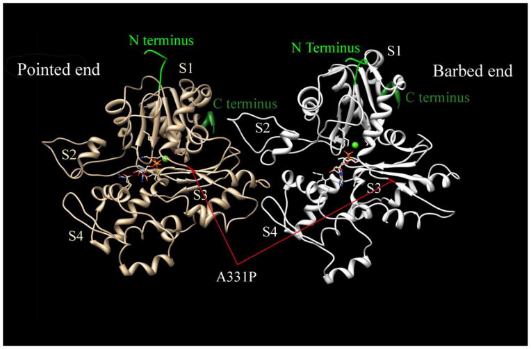
Figure 1. Actin dimer structure in F-form and the position of A331P mutation.
The N and C terminus of each actin monomer are labeled. S1–S4 represent each subdomain in the actin monomer. A331P mutation is marked in red in both actin monomers. Modified from [40](PDB ID:4a7f)

