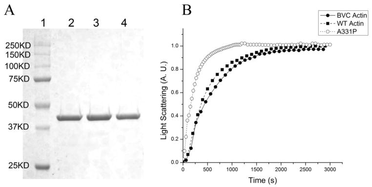
Figure 2. Purification and polymerization of recombinant actin.

(A) SDS-PAGE of purified actins. Lane 1: Precision plus protein dual color standards (Bio-rad); Lane 2: BVC actin; Lane 3: WT actin; Lane 4: A331P actin. (B) Polymerization of 0.2mg/ml G-actin, monitored as the increase of light scattering signal. All light scattering signals were normalized to the saturation level (at ~2800 s) of WT actin.
