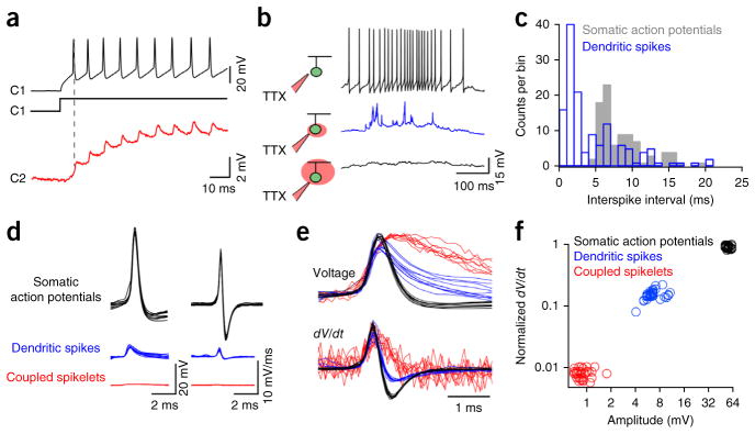
Figure 3.

Gap junction inputs on their own do not trigger dendritic spikes. (a) Spike trains evoked in C1 (black, 200-pA depolarizing current step injected through the patch electrode) drove coupled spikelets in C2 (red), which were exclusively mediated by gap junctions (spikelets are shown on a magnified scale). (b) Local application of TTX blocked somatic spikes (top, black), revealing dendritic spikes (middle, blue) that were abolished when TTX was applied over the entire cell (bottom). (c) A plot of the interspike interval (dendritic spikes (blue) and somatic action potentials (gray), n = 91 action potentials and 139 dendritic spikes from 4 cells) illustrates a ~5-ms refractory period for somatic action potentials, but not dendritic spikes. (d) Overlays of somatic action potentials (black), dendritic spikes (blue) and coupled spikelets (red), shown on the same scale (n = 10 events; left). The time derivative of these events is shown on the right. (e) Normalized versions of the traces shown in d, emphasizing the different kinetics of the somatically measured events. (f) The maximum rate of change in voltage plotted against the peak voltage for the three types of events (35 events are plotted for each type). For this plot, light-evoked somatic action potentials and dendritic spikes were from the same cell that was hyperpolarized to increase the failure of somatic action potentials (Supplementary Fig. 6), whereas coupled spikelets were measured using the protocol shown in a.
