
Figure 2.
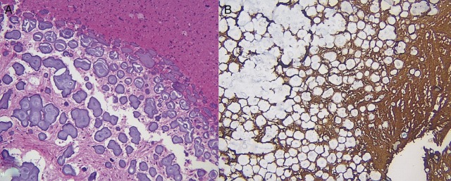
A well-circumscribed, extensively calcified lesion admixed with reactive astrocytosis. Multiple psammoma bodies are found on H&E staining (A). Immunohistochemistry is positive for glial fibrillar acidic protein (B) and negative for epithelial membrane antigen (not shown).