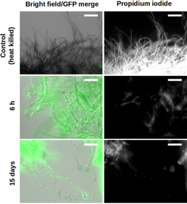
Fig 3.

Aspergillus niger remains alive when colonized by S. Typhimurium. Epifluorescence microscopy overlay images of live and heat-killed A. niger mycelia (filaments, grey) co-incubated with sfGFP-labelled S. Typhimurium cells (false coloured green, left panels) stained with propidium iodide (white, right panels). Scale bars: 50 μm.
