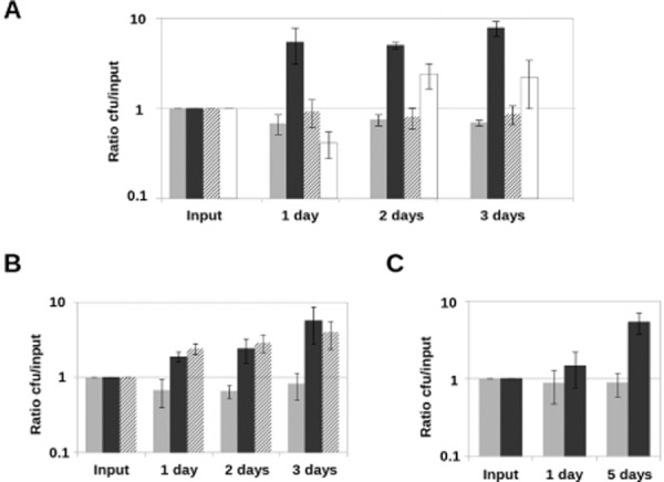
Fig 4.

Bacterial growth requires live fungi or dead fungal lysate. Bacteria were detached and quantified at different time points, and cfu were normalized with respect to input values.A. Light grey bars represent growth of bacteria alone, dark grey bars represent growth of S. Typhimurium in co-incubation with live A. niger, striped bars represent bacterial growth in co-incubation with heat-killed fungal filaments (washed after killing) and white bars represent growth in co-incubation with live fungi separated by a semipermeable membrane.B. Light grey bars represent growth of bacteria alone, dark grey bars represent growth of S. Typhimurium in co-incubation with live A. niger and striped bars represent bacterial growth in co-incubation with heat-killed fungi in the same potassium phosphate buffer where it was killed.C. Light grey bars represent growth of bacteria alone and dark grey ones represent growth of S. Typhimurium in presence of A. niger filtrate. All co-incubations were performed in potassium phosphate buffer. Error bars represent standard deviation of the values obtained in three independent biological replicates (n = 3). Y-axis values are represented in logarithmic scale.
