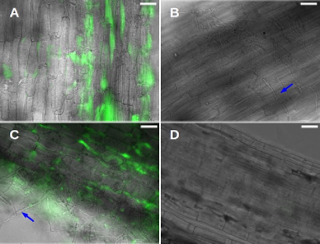
Fig 6.

Maize roots are colonized by S. Typhimurium and A. niger. Images are overlay of transmitted light (grey) with sfGFP fluorescence (false coloured green).A. Epidermal maize root tissue colonized by sfGFP-labelled S. Typhimurium.B. Epidermal maize root tissue colonized by A. niger (blue arrow).C. Epidermal maize root tissue colonized by sfGFP-labelled S. Typhimurium and A. niger. Blue arrow points at a representative fungal filament.D. Non-inoculated maize root tissue is shown as control. Scale bars: 50 μm.
