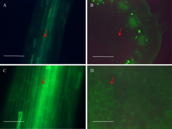
Fig 2.

Fluorescence micrographs showing the colonization of roots and stems of micropropagated Jerusalem artichoke seedlings by O. anthropi Mn1g. Arrows indicate visualized bacteria. (A, C) Endophytic colonization of tissues of surface-sterilized root at 15 DAI. (B, D) Endophytic colonization of surface-sterilized stem at 15 DAI. (A, B) Bar = 10 μm; (C, D) bar = 20 μm.
