Fig. 1.
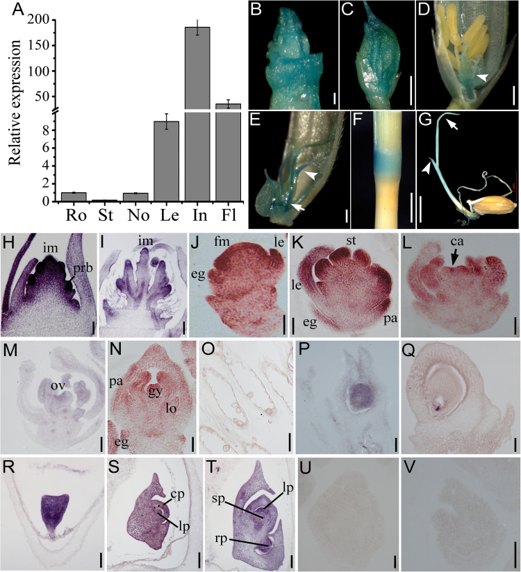
Expression patterns of OsIG1 in WT of rice. (A) Real-time quantitative RT-PCR analyses of OsIG1 expressions in various organs of WT rice. RNAs were collected from 3-week-old seedlings: root (Ro), shoot (St), leaf (Le) and inflorescence (In) at stage In5, with the node (No) and flower (Fl) at the mature embryo sac stage. Actin was used as a control. The expression of IG1 in root was set as 1. Error bars represent SD from three independent biological replicates. (B–G) Localization of GUS activity under the control of the OsIG1 promoter. (B) A 5 mm-long inflorescence; scale bar, 1mm. (C) A 3 mm-long spikelet; bar, 1mm. (D) Postmeiosis spikelet: arrowhead indicates ovary; bar, 200 µm. (E) The mature embryo sac stage: arrow and arrowhead indicate filament and style, respectively; bar, 200 µm. (F) Node; bar, 5mm. (G) Young seedling: arrow and arrowhead indicate primary leaf and second leaf, respectively; bar, 5mm. (H–V) In situ hybridization of cross-sections with an OsIG1-specifc antisense probe. (H, I) Longitudinal section of a young inflorescence. (J–N) Longitudinal sections of a developing WT spikelet. (O) Anther in nearly mature flowers. (P) Immature ovary. (Q) Mature ovary. (R–T) Sections of WT embryos at 2 days after pollination (DAP) (R), 5 DAP (S) and 7 DAP (T). (U, V) In situ analysis using a sense probe of OsIG1 in the early spikelet as negative controls. ca, carpel; cp, coleoptilar; eg, empty glume; fm, floral meristem; gy, gynoecium; im, inflorescence meristem; le, lemma; lo, lodicule; lp, leaf primordia; ov, ovule; pa, palea; prb, primary rachis branches; rp, root primordium; sp, shoot primordium; st, stamen. Scale bars, 50 μm in (H, J–O, R), 400 μm (I) and 100 μm in others.

