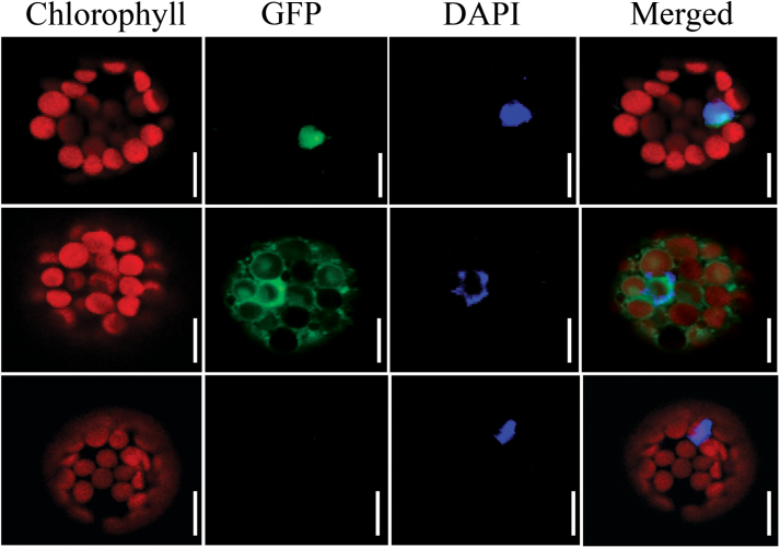
Fig. 2.
Localization of OsIG1-GFP fusion protein transiently expressed in Arabidopsis protoplasts. Upper panels, OsIG1-GFP; middle panels, 35S-GFP; bottom panels, an untransformed plant as a negative control. Left to right: red, chlorophyll autofluorescence; green, GFP fluorescence; blue, nucleus stained with DAPI; merged, combined fluorescence from GFP, chlorophyll and DAPI. Scale bars, 20 µm.

