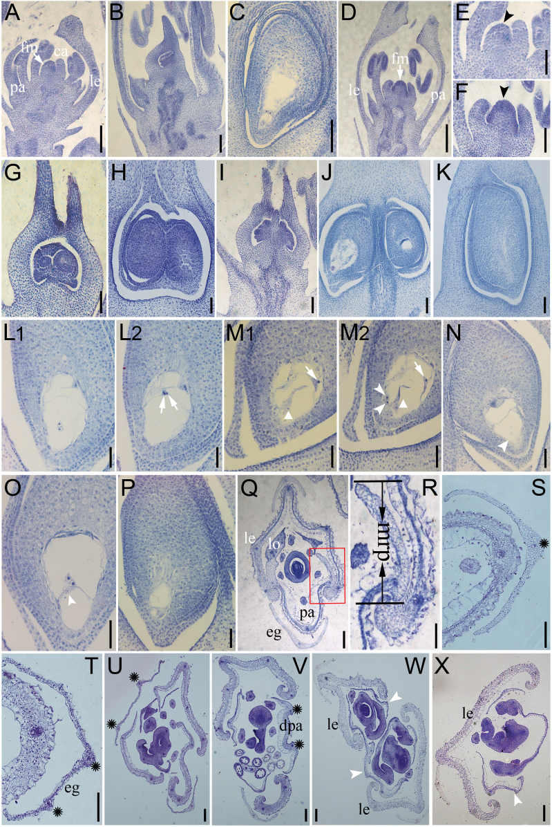
Fig. 4.
Histological analyses of WT and OsIG1-RNAi flowers. (A–C) Pistil development in the WT. A carpel protruded from the lemma side of the floral meristem (A). When the carpel completely enclosed the ovule primordium, a megaspore mother cell became visible and an integument primordium was differentiated in the ovule primordium (B). In a mature ovule, a vacuolated embryo sac was visible (C). (D) When a carpel protruded, the floral meristem in OsIG1-RNAi (D) was larger than that of the WT (A). (E, F) Close-up of floral meristem shown in the WT (A) and OsIG1-RNAi (D), respectively. (G–J) Unusual double ovules fused (G, H) or not fused (I, J) occurred in the transgenic ovaries. (K) Embryo sac degeneration in OsIG1-RNAi ovule. (L1, L2) Serial section showing OsIG1-RNAi embryo sac with extra polar nuclei (arrows). (M1, M2) Serial section showing OsIG1-RNAi embryo sac with possible multiple egg cells (arrowheads), extra synergids (triangular arrows) and polar nuclei located in an abnormal position close to the chalazal end (arrows). (N) Embryo sac without female germ unit (arrowhead). (O) Embryo sac with abnormal nuclear migration (or abnormal position of egg cell and synergid cells; arrowhead). (P) Abnormally small embryo sac. (Q–S) Cross-sections of a WT spikelet. (R) Magnification of the area within the box on Q showing interlocking of lemma and palea. (S) The WT empty glume with one vascular bundles (asterisk). (T-X) Cross-sections of the OsIG1-RNAi spikelets. (T, U) Two vascular bundles in the OsIG1-RNAi empty glumes (asterisks in T), as opposed to one in WT (asterisks in S). Edges of lemma and palea are hooked in the WT (Q), but there is no interlock with the lemma in the OsIG1-RNAi spikelet (U-X). (U) An abnormal flower with hull-like organs and two vascular bundles of empty glume (asterisks) in an OsIG1-RNAi flower. (V) The OsIG1-RNAi spikelet has only two full-developed vascular bundles (asterisks) in degenerated palea. (W) Transverse section of twin-flower spikelet (one spikelet containing two separate carpels) of OsIG1-RNAi; arrowheads indicate the degenerated palea. (X) The degenerated palea (arrowhead) lacking a bop and retaining mrp-like structures in an OsIG1-RNAi spikelet. ca, primary carpel; dpa, degenerated palea; eg, empty glume; fm, floral meristem; le, lemma; lo, lodicule; mrp, marginal region of palea; pa, palea. Scale bars, 100 μm (A–D, Q–X), 50 μm (E–P).

