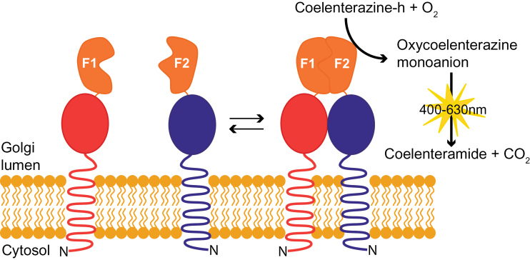
Fig. 1.
Schematic representation of the reversible Renilla luciferase protein complementation assay (Rluc-PCA) to study Golgi lumenal protein interactions. Membrane proteins with a type II membrane topology, spanning the membrane once with the N-terminus (N) in the cytosol and a lumenal C-terminus, are shown fused to the N-terminal domain (F1) and C-terminal domain (F2) of human-codon optimized Renilla luciferase (hRluc). Arrows denote the dynamics of the protein interaction, the coupling and decoupling of the two domains of hRluc. (This figure is available in colour at JXB online.)

