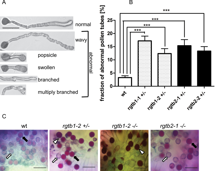
Fig. 3.
Male gametophyte morphology in Atrgtb mutants. (A) Morphological aberrations in pollen tubes germinated in vitro; scale bar=100 μm. (B) Frequency of pollen tube abnormalities; bars represent the mean ±SE. Statistical significance was analysed by Mann–Whitney test, ***P<0.001. (C) In vivo germination of mutant pollen on wt stigma. Alexander stain 16h after pollination; grey and pale pink pollen grains are germinated (arrows), dark purple grains are non-germinated (white arrowhead). Scale bar=100 μm. Note shrunken pollen grains deposited on stigma for Atrgtb1+/– mutant pollen (black arrowhead) and low frequency of germination for Atrgtb1–/– pollen. Representative images are shown.

