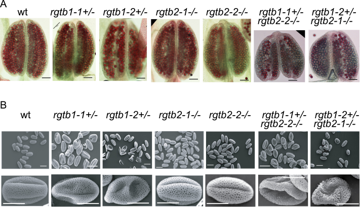
Fig. 6.
Pollen grain development in single mutants in AtRGTB and in the Atrgtb1Atrgtb2 double mutant. (A) Alexander stain of mature anthers; scale bar=100 μm. (B) SEM images of mature pollen grains. Note that in pollen derived from Atrgtb1+/– some grains are hollow, but of normal length, but in pollen derived from Atrgtb1+/–Atrgtb2–/–, half of the grains are shrunken and many of them show abnormal exine structure; scale bar=10 μm for higher magnification and 20 μm for lower magnification. Representative images are shown. (This figure is available in colour at JXB online.)

