
Figure 1.
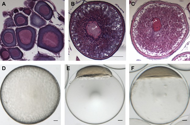
Overview of the developmental stages of Atlantic cod follicles, eggs, and embryos assessed with the microarray. Histological sections of pre-, early-, and late-vitellogenic follicles (A, B, and C, respectively) and photos of an unfertilized egg (D) and embryo at blastula stage (23.5 hr post-fertilization (hpf)) (E) and gastrula stage (58 hpf) (F). e, egg envelope; ca, cortical alveoli; n, nucleolus. Scale bar, 100 µm. [Color figure can be viewed in the online issue which is available at wileyonlinelibrary.com]