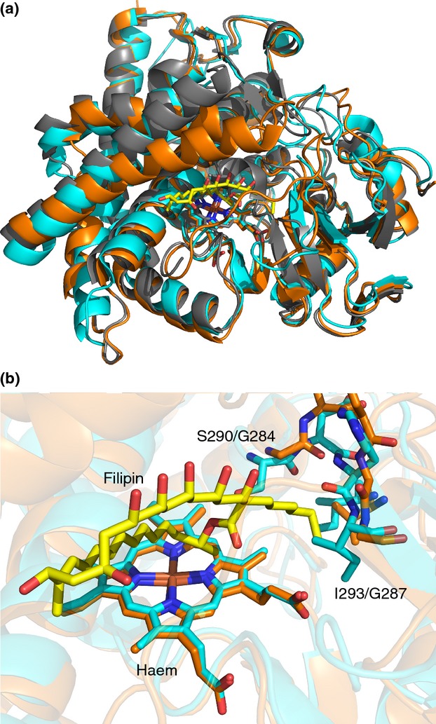
Figure 5.

Crystal structures of CYP105D6 and CYP105P1 from Streptomyces avermitilis. (a) Overlaid cartoon representations of CYP105D6 (( ) D6, PDB 3ABB; Xu et al. 2010), CYP105P1 ((
 ) P1, PDB 3E5J; Xu et al. 2009) and CYP105P1 with bound filipin I ((
 ) P1 + filipin, PDB 3ABA; Xu et al. 2010). Haem and filipin I (from PDB 3ABA) shown as sticks. (b) Active site regions of CYP105D6 and CYP105P1, with residues responsible for the differing substrate specificity of the two enzymes (see text), bound haem and filipin I shown as sticks.
