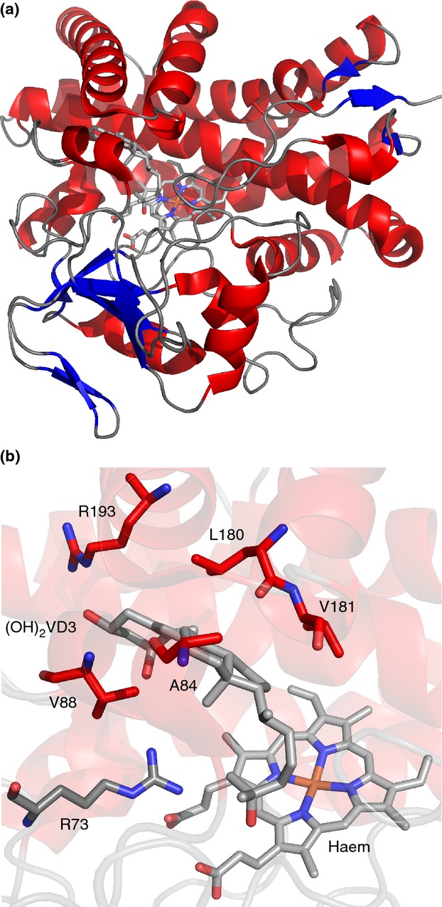
Figure 6.

Crystal structure of CYP105A1 from Streptomyces griseolus. (a) Cartoon representation of CYP105A1-R84A (PDB 2ZBZ, Sugimoto et al. 2008) with bound haem and 1α,25-dihydroxyvitamin D3 ((OH)2D3) shown as sticks. (b) Active site region of CYP105A1-R84A, with important residues (see text), bound haem and (OH)2D3 shown as sticks. The same colour scheme is used for the secondary structure elements as in Fig.4.
