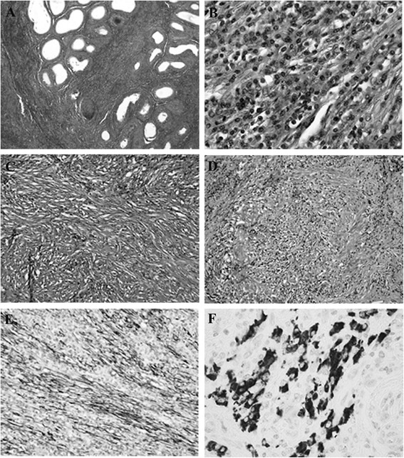
Figure 3.

Microscopic findings of surgical specimen. Hematoxylin-eosin staining of the paratesticular mass reveals widespread fibrosis and lymphocyte aggregation from the left epididymis to the spermatic cord (×40; A). Spindle cell proliferation with chronic inflammatory cells mostly comprised plasma cells with neither atypia nor mitosis (×200; B). Myofibroblastic cells with marked fibrosis reveals a storiform pattern (×100; C). Obliterative phlebitis is apparent (×100; D). For immunostaining, sections were stained with anti-alpha-smooth muscle actin (α-SMA) (1A4, 1:800, DAKO, Glostrup, Denmark) and -IgG4 (HP6025, 1:1280; ZYMED Laboratories, CA, USA) antibodies by using automated immunostainer (Ventana Benchmark, Tucson, AZ, USA). Spindle cells are positive for α-SMA, indicating myoepithelial cells (×100; E). More than 10 IgG4-positive plasma cells/HPF are seen on anti-IgG4 immunostaining (×400; F).
