Figure 1.

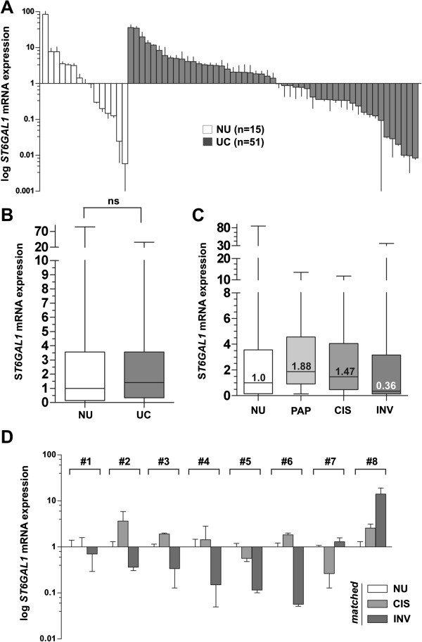
ST6GAL1 mRNA expression analyses in human bladder cancer. (A) Real-time PCR based ST6GAL1 mRNA expression analyses of 51 tumor samples (UC) compared to normal urothelium (NU) samples (n = 15). The median expression level of NU was set to 1. Vertical lines: ± standard error of margin (s.e.m.). (B) Box plot showing median ST6GAL1 mRNA expression in normal urothelium compared to all analyzed urothelial cancer samples. Horizontal lines: grouped medians. Boxes: 25–75% quartiles. Vertical lines: range, peak and minimum, ns: not significant. (C) Itemized box plot demonstrating median ST6GAL1 mRNA expression in normal urothelium (NU), non-invasive papillary tumors (PAP), CIS, and invasive bladder tumors (INV). Horizontal lines: grouped medians. Boxes: 25–75% quartiles. Vertical lines: range, peak and minimum. (D) Real-time PCR based ST6GAL1 mRNA expression analyses of patient triplets with matched normal urothelium, CIS and invasive tumor samples. Matched CIS and solid tumors were normalized to the corresponding NU, respectively. Vertical lines: + s.e.m.
