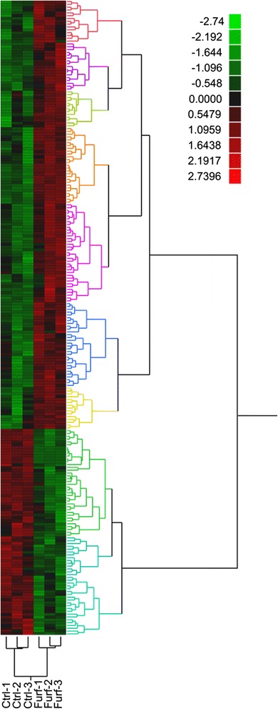
Figure 4.

Heat map of proteomic analysis with and without 15 mM furfural. Proteins exhibiting a statistically significant (ANOVA, P ≤0.05) difference in abundance are included. Each protein (row) was independently normalized to recast spectral count values as standard deviations from the row mean. Protein abundance differences were clustered according to trends measured across all biological replicates. Red = increased; green = decreased abundance.
