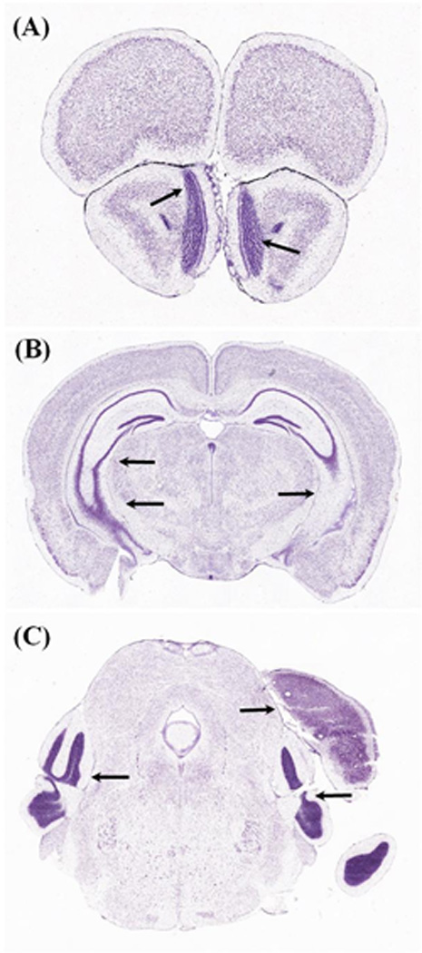
Figure 1. Three-examples of Misalignment in the Coronal Plane.

Obtaining whole brain sections, parallel to canonical planes of section, is an important but difficult goal in neurohistology. A–C, are three examples, from the widely used Allen Brain Atlas(Dong and Science, 2008), of A-P offset between the two hemispheres. This offset is due to tilting of the brain during cryostat positioning, which resulted in anterior shifting of the left hemisphere (and posterior shifting of the right hemisphere). This offset is indicated by black arrows, pointing to the olfactory bulb in A, hippocampus in B, and to the posterior cortices in C (left pointing arrows). The right pointing arrow in C, shows the posterior edge of the cortex (dorsal) and the hippocampus, which is present in the left-hemisphere, but missing from the right-hemisphere, at the same A-P level. Allen Reference Atlas, coronal level 27 is shown in (A); level 80 is shown in (B); and level 105 is shown in (C).
