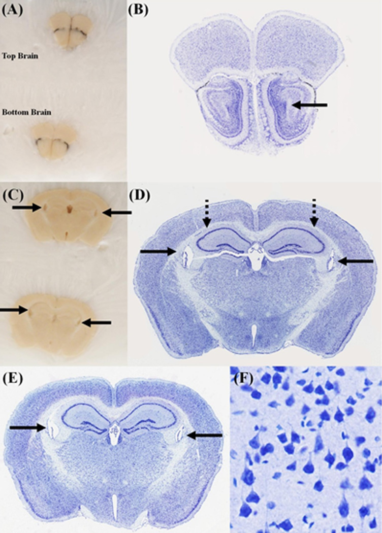
Figure 5. Coronal sections of two brains, processed using the Pre-Cast Mold System.

(A) and (C) Blockface images were taken at two coronal levels. The corresponding Nissl stained sections are shown in (B) and (D-F). The "top brain" is shown in (B) and (D). The bottom brain is shown in (E). The solid black arrows point to the lateral ventricles. The dotted black lines point to the Hippocampal formation (HPF). All sections appear symmetrical on the blockface, but a closer inspection of the stained sections, shows an offset of about 2–3 atlas plates. This is best seen in the lateral extent of the HPF. One of the advantages of this system, is that two brains can be frozen in the same block, and are cut at the same approximate level, as can be seen in (D) and (E). (F) A high-magnification view of the stained sections shows that the cellular morphology is well preserved.
