
Figure 1.
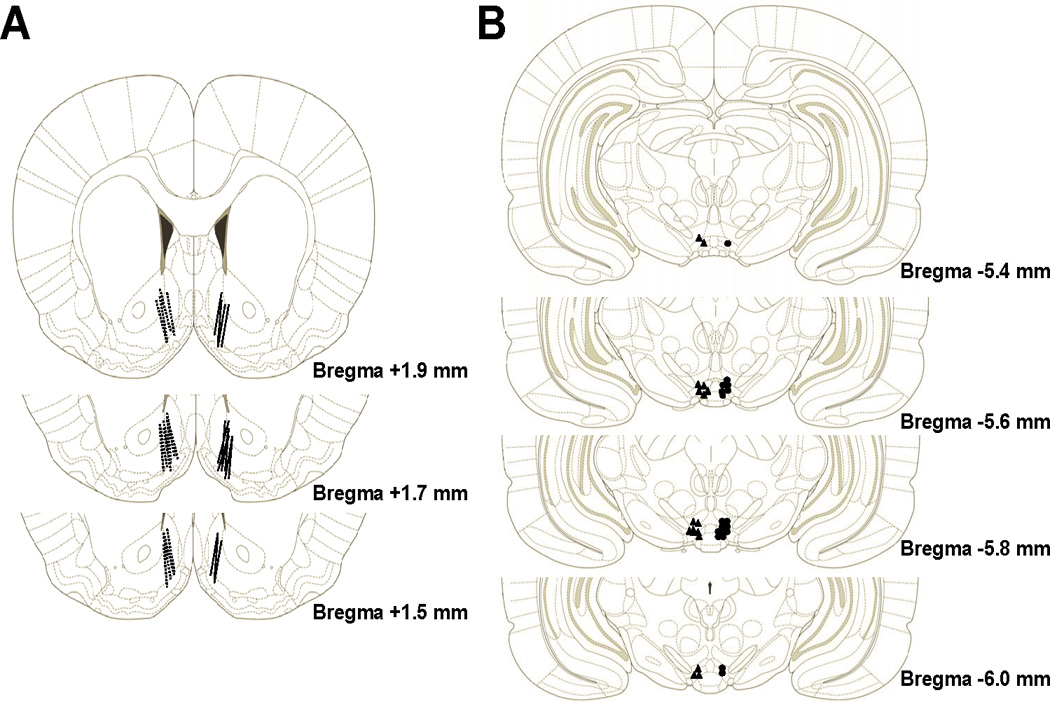
Represents the histological placements of the microdialysis cannula in the AcbSh (panel A) and the microinjection cannula in the posterior VTA (panel B) for Experiments 1 and 2. Placements for Experiment 1 are presented in the left hemisphere while placements for Experiment 2 are presented in the right hemisphere. Animals with cannula outside of these target brain regions were excluded from analyses. Histological plates have been adapted from Paxinos and Watson (2006).