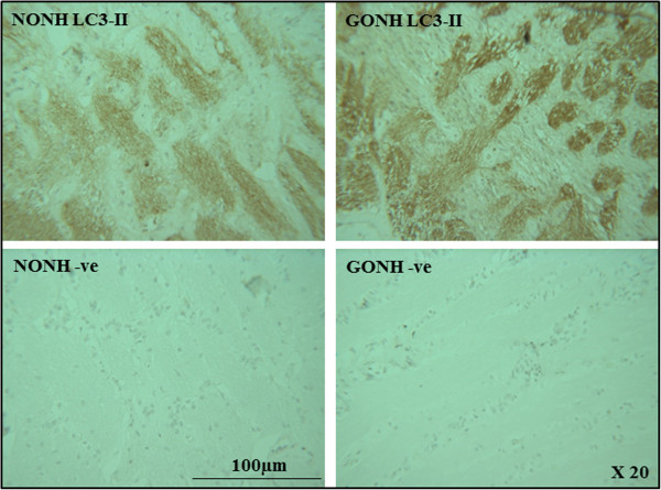
Figure 5.

Immunohistochemical staining of ONH sections with the autophagy marker LC3 II. Longitudinal sections through optic nerves from glaucomatous (GONH) and normal (NONH) donors at magnifications of 20X stained for LC3-II are shown. Cell nuclei stain purple-blue. Positive staining appears brown and is more abundant in that section from the donor with glaucoma.
