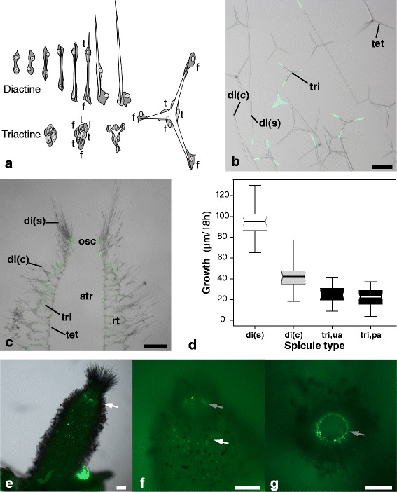
Figure 1.

Spicules and their formation in S. ciliatum . (a) Formation of diactines and triactines by sclerocytes in calcareous sponges (redrawn from [18,19]). (b) isolated spicules; scale bar: 100 μm. (c) skeletal arrangement; scale bar: 250 μm. (d) Spicule growth in 18 h observed in three spicule types of S. ciliatum. (e-g) Location of spicule formation in S. ciliatum (calcein disodium staining). Single spicules are formed all over the sponge body, with two regions of denser spicule formation: (1) the radial tube formation zone (white arrow, e) and the proximal tips of the slender diactines of the osculum (inside the osculum, grey arrow: f, g); scale bars: 250 μm. b,c,e: light microscopic images overlayed with fluorescence microscope images. f,g: fluorescence microscope images. Abbreviations: di(c) curved diactines from the distal end of the radial tubes; di(s): slender diactines of the oscular fringe; f: founder cell; t: thickener cell; tri: triactines; tet: tetractines.
