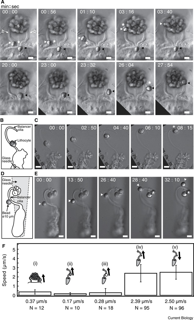
Figure 1.
Lithocyte and microsphere movements on balancer cilia. (A) Lithocyte transport along a pair of adjacent balancers of a 3 mm larva of Mnemiopsis leidyi, viewed in the sagittal body plane. Two refractile lithocytes bud off sequentially from the epithelial floor and move up each balancer (b) into the statolith (s). Upper row: the left lithocyte (white arrowhead) is liberated first and moves up the left balancer. White asterisks indicate the peripheral cytoplasm surrounding the refractile concretion. Lower row: the right lithocyte (black arrowhead) buds off later and moves up the right balancer. The white asterisk shows a leading cytoplasmic rim. Scale bar, 10 µm. (B) Schematic drawing of an isolated adult balancer and an attached lithocyte. The cell bodies of the balancer are held by a glass needle. A dissociated adult lithocyte on the coverslip was translocated to the balancer by microscope stage controls and positioned above the curved base of the balancer. (C) The lithocyte moves up the balancer toward the tip at a speed of ~0.14 µm/s. The cytoplasmic rim of the lithocyte is visible in the last frame (white asterisk). Scale bar, 10 µm. (D,E) 10 µm microsphere transport along an isolated adult balancer. (D) Schematic drawing of the isolated balancer with a 10 µm microsphere attached about halfway up the balancer above its curved base. Several lithocytes (L) from the statolith remain at the balancer tip. The cell bodies of the balancer are held with a glass needle (out of focus). (E) The 10 µm microsphere moves up the balancer toward the tip. Scale bar, 10 µm. (F) Speed and direction histogram of lithocyte and microsphere movements on balancers from different preparations. Arrows show directions of tipward (anterograde) and baseward (retrograde) movements. The numbers of analyzed displacements are indicated by N. (i) Freshly budded lithocytes on intact larval balancers (12 displacements of 5 lithocyte movements on 5 balancers were analyzed); (ii) dissociated adult lithocytes on isolated adult balancers (10 displacements of 3 dissociated lithocyte movements on 3 isolated balancers were analyzed); (iii) 10 µm microspheres on isolated adult balancers (18 displacements of 7 microsphere movements on 7 isolated balancers were analyzed); (iv,v) 1 µm microspheres on isolated adult balancers (191 displacements of 11 microsphere movements on 10 isolated balancers were analyzed).

